| Home | |
| Faculty | |
| Graduate Program | |
| Events and Seminars | |
| Resources | |
| Contact Us | |
|
F A C U L T Y P R O F I L E
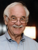
Current Research Understanding the function of molecular receptors has been my guiding interest and the central mission of the Center. Receptors function in three steps: signal recognition, transduction, and transmission. This is exemplified by the nicotinic acetylcholine receptors, on which I worked for forty years. These receptors bind acetylcholine, transduce this binding into the opening of a pore, and conduct cations causing a depolarization of the synaptic membrane. We identified the overall receptor structure and, within this, the specific sites and substructures at the front-line of these functional steps. More recently, this approach was applied to the BK channel, a voltage- and calcium-ion-activated channel, widely expressed and responsible for membrane hyperpolarization. Also recently, I began to consider cellular functions that depend on circuits of signaling components. In these cases, in addition to individual molecular structures, cellular structures and the distribution of the molecular components in these structures determine the nature of the signaling that underlies the cellular function. Crucial to the specificity and effectiveness of the signaling are microdomains, which both channel and amplify the signals. My approach to understanding such systems is to represent all steps as kinetic equations and to solve the resulting equations in time and space. To the extent that experimental data exist, the equations are fit to these data. Such a model is a mathematical summary of the known properties of the components and of the target cellular function. It is also kinetically plausible and consistent with thermodynamics. Notwithstanding, such a model is a hypothetical outline of how the cell might function, always wanting experimental tests and refinement.
Selected Publications Karlin, A. 2002. Emerging structure of the nicotinic acetylcholine receptors. Nature Reviews Neuroscience 3: 102-114. Liu, G., Zakharov, S., Yang., L., Deng, S., Landry, D., Karlin, A., Marx, S. (2008) Position and role of the BK channel alpha subunit S0 helix inferred from disulfide crosslinking. J. Gen. Physiol 131: 537-548. Liu, G., Zakharov, S., Yang., L., Wu, R., Deng, S., Landry, D., Karlin, A., Marx, S. (2008) Locations of the beta1 transmembrane helices in the BK potassium channel. Proc. Natl. Acad. Sci. USA 105:10727-10732. Chung, D.Y., Chan, P. J., Bankston, J.R., Yang, L., Liu, G., Marx, S.O., Karlin, A., Kass, R.S. (2008) Location of KCNE1 relative to KCNQ1 in the IKS potassium channel by disulfide crosslinking of substituted cysteines. Proc. Natl. Acad. Sci. USA 106:743-748. Wu, R.S., Chudasama, N., Zakharov, S.I., Doshi, D., Motoike, H., Liu, G., Yao, Y., Niu, X., Deng, S.-X., Landry, D.W., Karlin, A., and Marx, S.O. (2009) Location of the beta4 transmembrane helices in the BK potassium channel. J. Neurosci. 29:8321-8328. PMCID:PMC2737688 Liu, G., Niu, X., Wu, R.S., Chudasama, N., Yao, Y., Jin, X., Weinberg, R., Zakharov, S.I., Motoike, H., Marx, S.O., and Karlin, A. (2010) Location of modulatory β subunits in BK potassium channels. J. Gen. Physiol. 135:449-459; PMCID: PMC2860586 Chan, P.J., Osteen, J.D., Xiong, D., Bohnen, M.S., Doshi, D., Sampson, K.J., Marx, S.O., Karlin, A., and Kass, R.S. (2012) Characterization of KCNQ1 atrial fibrillation mutations reveals distinct dependence on KCNE1. J. Gen Physiol. 139:135-144 PMCID: PMC3269792 Wu RS, Liu G, Zakharov SI, Chudasama N, Motoike H, Karlin A, Marx SO. (2013 ) Positions of β2 and β3 subunits in the large-conductance calcium- and voltage-activated BK potassium channel. J Gen Physiol. 141:105-17. PMCID:PMC3536527 Niu X, Liu G, Wu RS, Chudasama N, Zakharov SI, et al. (2013) Orientations and proximities of the extracellular ends of transmembrane helices S0 and S4 in open and closed BK potassium channels. PLoS ONE 8(3): e58335. doi:10.1371/journal.pone.0058335 PMCID: PMC3589268 Liu G, Zakharov SI, Yao Y, Marx SO, and Karlin, A (2015) Positions of the cytoplasmic end of BK alpha S0 helix relative to S1-S6 and of beta1 TM1 and TM2 relative to S0-S6. J Gen Physiol 145:185-199. doi:10.1085/jgp.201411337. PMCID:PMC4338161 Karlin A (2015) Membrane potential and Ca2+ concentration dependence on pressure and vasoactive agents in arterial smooth muscle: A model. J. Gen. Physiol. 146:79–96 www.jgp.org/cgi/doi/10.1085/jgp.201511380. PMCID:PMC4485026
|

